Video Library

Introduction to Chest Radiography
Explanation of the anatomy of the mediastinum and a systematic approach to reading chest radiographs.
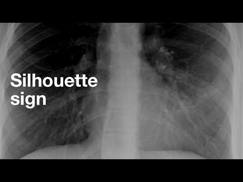
The Silhouette Sign
Textbooks I like for chest radiology— Med students and all residents: Felson’s Principles of Chest Roentgenology https://amzn.to/3FhBkvN Radiology residents: Thoracic Imaging: Pulmonary and Cardiovascular Radiology https://amzn.to/2YqzLLh Thoracic radiology fellows: Muller’s Imaging of the Chest: Expert Radiology Series https://amzn.to/3ouJ7QY

The Pulmonary Hila
Textbooks I like for chest radiology— Med students and all residents: Felson’s Principles of Chest Roentgenology https://amzn.to/3FhBkvN Radiology residents: Thoracic Imaging: Pulmonary and Cardiovascular Radiology https://amzn.to/2YqzLLh Thoracic radiology fellows: Muller’s Imaging of the Chest: Expert Radiology Series https://amzn.to/3ouJ7QY
Pulmonary Edema
Textbooks I like for chest radiology— Med students and all residents: Felson’s Principles of Chest Roentgenology https://amzn.to/3FhBkvN Radiology residents: Thoracic Imaging: Pulmonary and Cardiovascular Radiology https://amzn.to/2YqzLLh Thoracic radiology fellows: Muller’s Imaging of the Chest: Expert Radiology Series https://amzn.to/3ouJ7QY

Community Acquired Pneumonia
The appearance of community acquired pneumonia on CXR. Link to the Google Drive Presentation: https://docs.google.com/presentation/d/1mQaP0-jSG6Dj9LPHk6L5913Lu7yTlVEZjK_t8f9xELg/edit?usp=sharing

ET Tubes on Chest X-ray
Quick tutorial on how to evaluate ET tubes on chest radiography.

The Diaphragm on the Lateral CXR
Here are the 5 ways to distinguish which side of the diaphragm is which on the lateral CXR: 1. The stomach bubble and splenic flexure of the colon are below the left. 2. The right hemidiaphragm is usually higher than the left. 3. The left hemidiaphragm is obscured by the heart anteriorly. 4. The right hemidiaphragm terminates posteriorly by the larger right ribs., 5. Look for pathology.

How I Read a Chest CT
This is for first year residents, starting out on their chest rotation. This is the order in which I read a chest CT. When you first start out, it is important to stick to a script so that you read studies the same way each time. It may be beneficial to have a checklist that you follow to make sure you aren't missing anything. Textbooks I like for chest radiology— Med students and all residents: Felson’s Principles of Chest Roentgenology https://amzn.to/3FhBkvN Radiology residents: Thoracic Imaging: Pulmonary and Cardiovascular Radiology https://amzn.to/2YqzLLh Thoracic radiology fellows: Muller’s Imaging of the Chest: Expert Radiology Series https://amzn.to/3ouJ7QY


Lobar and Segmental Lung Anatomy on CT
How to find the lobar and segmental bronchi on axial CT. This video is meant for radiology residents, but would benefit thoracic surgery residents and pulmonary medicine fellows as well. Textbooks I like for chest radiology— Med students and all residents: Felson’s Principles of Chest Roentgenology https://amzn.to/3FhBkvN Radiology residents: Thoracic Imaging: Pulmonary and Cardiovascular Radiology https://amzn.to/2YqzLLh Thoracic radiology fellows: Muller’s Imaging of the Chest: Expert Radiology Series https://amzn.to/3ouJ7QY

The Lymph Node Stations in the Chest
Check out the IASLC lymph node map here: https://www.iaslc.org/Portals/0/35348-cards-erx_combined_trap_card2_copy.pdf?ver=2019-05-22-154420-740 Check out the Radiographics article I mentioned here: https://pubs.rsna.org/doi/full/10.1148/rg.346130097 Textbooks I like for chest radiology— Med students and all residents: Felson’s Principles of Chest Roentgenology https://amzn.to/3FhBkvN Radiology residents: Thoracic Imaging: Pulmonary and Cardiovascular Radiology https://amzn.to/2YqzLLh Thoracic radiology fellows: Muller’s Imaging of the Chest: Expert Radiology Series https://amzn.to/3ouJ7QY

The Secondary Pulmonary Lobule
Short description of the anatomy of the secondary pulmonary lobule. Check out this article for more information on this topic: https://www.ncbi.nlm.nih.gov/pubmed/16543587 Textbooks I like for chest radiology— Med students and all residents: Felson’s Principles of Chest Roentgenology https://amzn.to/3FhBkvN Radiology residents: Thoracic Imaging: Pulmonary and Cardiovascular Radiology https://amzn.to/2YqzLLh Thoracic radiology fellows: Muller’s Imaging of the Chest: Expert Radiology Series https://amzn.to/3ouJ7QY

Pulmonary CT Angiogram Basics
Textbooks I like for chest radiology— Med students and all residents: Felson’s Principles of Chest Roentgenology https://amzn.to/3FhBkvN Radiology residents: Thoracic Imaging: Pulmonary and Cardiovascular Radiology https://amzn.to/2YqzLLh Thoracic radiology fellows: Muller’s Imaging of the Chest: Expert Radiology Series https://amzn.to/3ouJ7QY

Algorithmic Approach to Multiple Lung Nodules
Great article: https://www.ncbi.nlm.nih.gov/pubmed/16537886 Textbooks I like for chest radiology— Med students and all residents: Felson’s Principles of Chest Roentgenology https://amzn.to/3FhBkvN Radiology residents: Thoracic Imaging: Pulmonary and Cardiovascular Radiology https://amzn.to/2YqzLLh Thoracic radiology fellows: Muller’s Imaging of the Chest: Expert Radiology Series https://amzn.to/3ouJ7QY

Chronic Consolidation
Acute consolidation has a pretty short differential. When a consolidation becomes chronic, you have to widen your differential. This video will help you form a short but powerful differential for a chronic consolidation with the mnemonic SPACE-V. Here are my references which I highly recommend you check out: https://pubmed.ncbi.nlm.nih.gov/31200867/ https://pubmed.ncbi.nlm.nih.gov/18716117/ https://pubmed.ncbi.nlm.nih.gov/17495282/ https://pubmed.ncbi.nlm.nih.gov/20413748/ https://www.flickr.com/photos/pulmonary_pathology/

4 Must-Know Features of ILD
Here are 4 findings you have to recognize and classify to understand ILD. Ground glass opacity Reticulation Traction Bronchiectasis Honeycombing Here's the link to the Fleischner Glossary: https://pubs.rsna.org/doi/10.1148/radiol.2462070712 Textbooks I like for chest radiology— Med students and all residents: Felson’s Principles of Chest Roentgenology https://amzn.to/3FhBkvN Radiology residents: Thoracic Imaging: Pulmonary and Cardiovascular Radiology https://amzn.to/2YqzLLh Thoracic radiology fellows: Muller’s Imaging of the Chest: Expert Radiology Series https://amzn.to/3ouJ7QY


Subsolid Nodules
This is a description of subsolid nodules, what they are and why we treat them differently from solid nodules. Here are some of the references I used in this article: The revised lung adenocarcinoma classification—an imaging guide https://www.ncbi.nlm.nih.gov/pmc/articles/PMC4209391/ CT screening for lung cancer: frequency and significance of part-solid and nonsolid nodules https://www.ajronline.org/doi/full/10.2214/ajr.178.5.1781053 Guidelines for Management of Incidental Pulmonary Nodules Detected on CT Images: From the Fleischner Society 2017 https://pubs.rsna.org/doi/10.1148/radiol.2017161659 LungRADS 1.1 https://www.acr.org/-/media/ACR/Files/RADS/Lung-RADS/LungRADSAssessmentCategoriesv1-1.pdf=la=en

Board Review | Thoracic Radiology | Part 1
Board review for thoracic radiology. Textbooks I like for chest radiology— Med students and all residents: Felson’s Principles of Chest Roentgenology https://amzn.to/3FhBkvN Radiology residents: Thoracic Imaging: Pulmonary and Cardiovascular Radiology https://amzn.to/2YqzLLh Thoracic radiology fellows: Muller’s Imaging of the Chest: Expert Radiology Series https://amzn.to/3ouJ7QY View my slides here: https://docs.google.com/presentation/d/1aRlsYsQN0z-EnczFofIw7oGxJmAVCyb4oPuIv6fCeFQ/edit?usp=sharing

Board Review | Thoracic Radiology | Part 2
Textbooks I like for chest radiology— Med students and all residents: Felson’s Principles of Chest Roentgenology https://amzn.to/3FhBkvN Radiology residents: Thoracic Imaging: Pulmonary and Cardiovascular Radiology https://amzn.to/2YqzLLh Thoracic radiology fellows: Muller’s Imaging of the Chest: Expert Radiology Series https://amzn.to/3ouJ7QY

Board Review | Thoracic Radiology | Part 3
Textbooks I like for chest radiology— Med students and all residents: Felson’s Principles of Chest Roentgenology https://amzn.to/3FhBkvN Radiology residents: Thoracic Imaging: Pulmonary and Cardiovascular Radiology https://amzn.to/2YqzLLh Thoracic radiology fellows: Muller’s Imaging of the Chest: Expert Radiology Series https://amzn.to/3ouJ7QY

Board Review | Thoracic Radiology | Part 4
Textbooks I like for chest radiology— Med students and all residents: Felson’s Principles of Chest Roentgenology https://amzn.to/3FhBkvN Radiology residents: Thoracic Imaging: Pulmonary and Cardiovascular Radiology https://amzn.to/2YqzLLh Thoracic radiology fellows: Muller’s Imaging of the Chest: Expert Radiology Series https://amzn.to/3ouJ7QY Cases: 00:00 Case 31 00:18 Case 32 00:30 Case 33 00:42 Case 34 01:04 Case 35 01:26 Case 36 01:59 Case 37 02:20 Case 38 02:32 Case 39 02:54 Case 40 03:17 Case 31 Answer 04:05 Case 32 Answer 05:02 Case 33 Answer 05:48 Case 34 Answer 06:57 Case 35 Answer 08:54 Case 36 Answer 10:37 Case 37 Answer 11:29 Case 38 Answer 12:13 Case 39 Answer 14:35 Case 40 Answer

CT signs of right heart strain with PE
This video goes over the CT signs of right heart strain that can be seen with acute pulmonary embolism. I describe and show examples of RV enlargement, septal bowing/flattening, pulmonary artery enlargement, and reflux of contrast into the IVC.

Upper Lung Predominant Fibrosis
Here's my differential for upper lung predominant pulmonary fibrosis: SHORTI: Sarcoid Hypersensitivity pneumonitis (chronic) Occupational pneumoconioses (silicosis, coal worker's, berylliosis) Radiation fibrosis TB and fungus Idiopathic pleuroparenchymal fibroelastosis Textbooks I like for chest radiology— Med students and all residents: Felson’s Principles of Chest Roentgenology https://amzn.to/3FhBkvN Radiology residents: Thoracic Imaging: Pulmonary and Cardiovascular Radiology https://amzn.to/2YqzLLh Thoracic radiology fellows: Muller’s Imaging of the Chest: Expert Radiology Series https://amzn.to/3ouJ7QY

A Better Approach to Studying for Radiology
In this video, I talk about a different way to study. It is centered around the idea of a mental checklist based on the clinical history of the patient.

Pneumothorax vs. Skin Fold
Here's how you can always tell the difference between a pneumothorax and a skin fold. Don't make this mistake!

Pneumothorax vs Lung Collapse
Just a quick video explaining the difference between the terms lung collapse and pneumothorax. Textbooks I like for chest radiology— Med students and all residents: Felson’s Principles of Chest Roentgenology https://amzn.to/3FhBkvN Radiology residents: Thoracic Imaging: Pulmonary and Cardiovascular Radiology https://amzn.to/2YqzLLh Thoracic radiology fellows: Muller’s Imaging of the Chest: Expert Radiology Series https://amzn.to/3ouJ7QY 00:00 Introduction 00:28 Definition of terms 01:53 Lung collapse without pneumothorax 02:53 Pleural effusion without pneumothorax 03:46 Collapse with pneumothorax 04:13 Pneumothorax without collapse 04:31 Summary

Central Lines on CXR
In this video, I go over how to tell the position of a central line. The central venous anatomy and landmarks are reviewed. Common positions of malpositioned lines are reviewed. Also, since more than 60% of you are viewing on mobile, I decided to make this a vertical video. Let me know how you like it. Textbooks I like for chest radiology— Med students and all residents: Felson’s Principles of Chest Roentgenology https://amzn.to/3FhBkvN Radiology residents: Thoracic Imaging: Pulmonary and Cardiovascular Radiology https://amzn.to/2YqzLLh Thoracic radiology fellows: Muller’s Imaging of the Chest: Expert Radiology Series https://amzn.to/3ouJ7QY

Pneumomediastinum
The radiographic appearance of pneumomediastinum and common clinical scenarios where you will see pneumomediastinum. Two articles mentioned in this video: Incidence, risk factors and outcome of barotrauma in mechanically ventilated patients https://pubmed.ncbi.nlm.nih.gov/14991090/ Pneumomediastinum and subcutaneous emphysema in COVID-19: barotrauma or lung frailty? https://pubmed.ncbi.nlm.nih.gov/33257914/

3 Common Patterns of Radiation Fibrosis (not from lung cancer)
In this video, I go over 3 patterns of radiation fibrosis that you will commonly see in the chest, but not from lung cancer. 1) Mediastinal radiation 2) Breast radiation 3) Head and neck radiation Chapters: 00:00 Introduction 00:19 Mediastinal radiation 06:46 Breast radiation 10:28 Head and neck radiation Textbooks I like for chest radiology— Med students and all residents: Felson’s Principles of Chest Roentgenology https://amzn.to/3FhBkvN Radiology residents: Thoracic Imaging: Pulmonary and Cardiovascular Radiology https://amzn.to/2YqzLLh Thoracic radiology fellows: Muller’s Imaging of the Chest: Expert Radiology Series https://amzn.to/3ouJ7QY

ILD: Organizing the Findings
The best place to start with an ILD case is to figure out the distribution - both in the craniocaudal and axial distribution. Textbooks I like for chest radiology— Med students and all residents: Felson’s Principles of Chest Roentgenology https://amzn.to/3FhBkvN Radiology residents: Thoracic Imaging: Pulmonary and Cardiovascular Radiology https://amzn.to/2YqzLLh Thoracic radiology fellows: Muller’s Imaging of the Chest: Expert Radiology Series https://amzn.to/3ouJ7QY 00:00 Introduction 00:15 Roadmap 00:42 Craniocaudal distribution 01:48 Upper lung disease 03:06 Lower lung disease 04:29 Axial distribution 05:37 Describe the findings 06:30 Pattern vs diagnosis 07:52 Conclusion

How I Read a Lateral CXR
Hi everybody. In this video, I show you how I look at a lateral CXR. I'm not going to do an exhaustive inventory of the anatomy in this video, but stay tuned for that in the future. Textbooks I like for chest radiology— Med students and all residents: Felson’s Principles of Chest Roentgenology https://amzn.to/3FhBkvN Radiology residents: Thoracic Imaging: Pulmonary and Cardiovascular Radiology https://amzn.to/2YqzLLh Thoracic radiology fellows: Muller’s Imaging of the Chest: Expert Radiology Series https://amzn.to/3ouJ7QY

What's the deal with MIPs?
In this video, I talk about what MIP images are and how they're useful for imaging the lungs. Textbooks I like for chest radiology— Med students and all residents: Felson’s Principles of Chest Roentgenology https://amzn.to/3FhBkvN Radiology residents: Thoracic Imaging: Pulmonary and Cardiovascular Radiology https://amzn.to/2YqzLLh Thoracic radiology fellows: Muller’s Imaging of the Chest: Expert Radiology Series https://amzn.to/3ouJ7QY

Lobar Collapse (examples of each lobe)
This video is all about lobar collapse or lobar atelectasis on chest radiographs. These are must-know patterns for radiologists, pulmonologists, and anyone else who works in critical care. Textbooks I like for chest radiology— Med students and all residents: Felson’s Principles of Chest Roentgenology https://amzn.to/3FhBkvN Radiology residents: Thoracic Imaging: Pulmonary and Cardiovascular Radiology https://amzn.to/2YqzLLh Thoracic radiology fellows: Muller’s Imaging of the Chest: Expert Radiology Series https://amzn.to/3ouJ7QY 00:00 Introduction 00:24 Right upper lobe collapse 02:37 Right middle lobe collapse 04:00 Right lower lobe collapse 06:09 Right middle and lower lobe collapse 07:50 Left upper lobe collapse 09:27 Left lower lobe collapse

3 Minute Papers: Lobectomy vs Segmentectomy for Lung Cancer
In this video, I summarize a recently published paper in the Lancet entitled: Segmentectomy versus lobectomy in small-sized peripheral non-small-cell lung cancer (JCOG0802/WJOG4607L): a multicentre, open-label, phase 3, randomised, controlled, non-inferiority trial" Here's a link to the pub med abstract: https://pubmed.ncbi.nlm.nih.gov/35461558/ Check out my video on how to localize the segmental bronchi on chest CT: https://youtu.be/HHYzFgJ7sjU Textbooks I like for chest radiology— Med students and all residents: Felson’s Principles of Chest Roentgenology https://amzn.to/3FhBkvN Radiology residents: Thoracic Imaging: Pulmonary and Cardiovascular Radiology https://amzn.to/2YqzLLh Thoracic radiology fellows: Muller’s Imaging of the Chest: Expert Radiology Series https://amzn.to/3ouJ7QY

Counting Ribs
Here's how to count ribs on chest x-ray. Start with the anterior aspect of the first rib and follow it back. That's your starting point. The next rib down is the posterior aspect of the second rib, and so on. Textbooks I like for chest radiology (Amazon links) — Med students and all residents: Felson’s Principles of Chest Roentgenology https://amzn.to/3FhBkvN Radiology residents: Thoracic Imaging: Pulmonary and Cardiovascular Radiology https://amzn.to/2YqzLLh Thoracic radiology fellows: Muller’s Imaging of the Chest: Expert Radiology Series https://amzn.to/3ouJ7QY #shorts
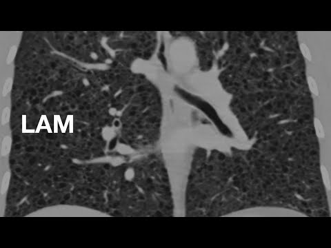
Lymphangioleiomyomatosis (LAM)
In this video, I cover the cystic lung disease, lymphangioleiomyomatosis (LAM). There are a few important clinical practice guideline articles that are very useful to both clinicians and radiologists which can be viewed here: https://www.thoracic.org/statements/guideline-implementation-tools/lam-diagnosis-and-mgmt.php Textbooks I like for chest radiology— Med students and all residents: Felson’s Principles of Chest Roentgenology https://amzn.to/3FhBkvN Radiology residents: Thoracic Imaging: Pulmonary and Cardiovascular Radiology https://amzn.to/2YqzLLh Thoracic radiology fellows: Muller’s Imaging of the Chest: Expert Radiology Series https://amzn.to/3ouJ7QY

Hampton's Hump
A Hampton's hump is a peripheral, wedge-shaped opacity on chest x-ray that represents a pulmonary infarct. Patient's frequently complain of pleuritic chest pain. If you see that combination of peripheral opacity and chest pain, think about pulmonary infarct and get a CT pulmonary angiogram. Textbooks I like for chest radiology (Amazon Links) — Med students and all residents: Felson’s Principles of Chest Roentgenology https://amzn.to/3FhBkvN Radiology residents: Thoracic Imaging: Pulmonary and Cardiovascular Radiology https://amzn.to/2YqzLLh Thoracic radiology fellows: Muller’s Imaging of the Chest: Expert Radiology Series https://amzn.to/3ouJ7QY #shorts

Lung Cancer Staging (Introduction)
This is a general overview of lung cancer staging before we discuss each of the T, N, and M categories which will each be separate videos. Textbooks I like for chest radiology— Med students and all residents: Felson’s Principles of Chest Roentgenology https://amzn.to/3FhBkvN Radiology residents: Thoracic Imaging: Pulmonary and Cardiovascular Radiology https://amzn.to/2YqzLLh Thoracic radiology fellows: Muller’s Imaging of the Chest: Expert Radiology Series https://amzn.to/3ouJ7QY

Lung Cancer Staging - Tumor (T)
Detailed review of the T category for TNM lung cancer staging (8th edition) Textbooks I like for chest radiology— Med students and all residents: Felson’s Principles of Chest Roentgenology https://amzn.to/3FhBkvN Radiology residents: Thoracic Imaging: Pulmonary and Cardiovascular Radiology https://amzn.to/2YqzLLh Thoracic radiology fellows: Muller’s Imaging of the Chest: Expert Radiology Series https://amzn.to/3ouJ7QY

Lung Cancer Staging - Node (N)
Detailed review of the N category for TNM lung cancer staging (8th edition) Textbooks I like for chest radiology— Med students and all residents: Felson’s Principles of Chest Roentgenology https://amzn.to/3FhBkvN Radiology residents: Thoracic Imaging: Pulmonary and Cardiovascular Radiology https://amzn.to/2YqzLLh Thoracic radiology fellows: Muller’s Imaging of the Chest: Expert Radiology Series https://amzn.to/3ouJ7QY
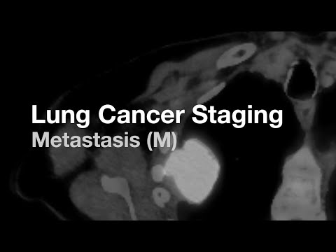
Lung Cancer Staging - Metastasis (M)
Detailed review of the M category for TNM lung cancer staging (8th edition) Textbooks I like for chest radiology— Med students and all residents: Felson’s Principles of Chest Roentgenology https://amzn.to/3FhBkvN Radiology residents: Thoracic Imaging: Pulmonary and Cardiovascular Radiology https://amzn.to/2YqzLLh Thoracic radiology fellows: Muller’s Imaging of the Chest: Expert Radiology Series https://amzn.to/3ouJ7QY

Suboptimal Studies
As the radiologist, we are the ultimate judge of image quality. It is up to you to have a discussion with the technologist if a study is not adequate.

Pseudoplaques
Quick explainer on pseudoplaques. They look like pleural plaques, but they aren't. They are in the lung. Usually seen in sarcoid. Textbooks I like for chest radiology— Med students and all residents: Felson’s Principles of Chest Roentgenology https://amzn.to/3FhBkvN Radiology residents: Thoracic Imaging: Pulmonary and Cardiovascular Radiology https://amzn.to/2YqzLLh Thoracic radiology fellows: Muller’s Imaging of the Chest: Expert Radiology Series https://amzn.to/3ouJ7QY

Asbestos-Related Pleural Plaques
In this video, I cover the CXR and CT appearance of asbestos-related calcified pleural plaques. Textbooks I like for chest radiology— Med students and all residents: Felson’s Principles of Chest Roentgenology https://amzn.to/3FhBkvN Radiology residents: Thoracic Imaging: Pulmonary and Cardiovascular Radiology https://amzn.to/2YqzLLh Thoracic radiology fellows: Muller’s Imaging of the Chest: Expert Radiology Series https://amzn.to/3ouJ7QY

Normal CT Appearance of a Lobectomy
In this video, I cover the normal appearance of each lobectomy and the resulting configuration of the remaining lobes. Textbooks I like for chest radiology— Med students and all residents: Felson’s Principles of Chest Roentgenology https://amzn.to/3FhBkvN Radiology residents: Thoracic Imaging: Pulmonary and Cardiovascular Radiology https://amzn.to/2YqzLLh Thoracic radiology fellows: Muller’s Imaging of the Chest: Expert Radiology Series https://amzn.to/3ouJ7QY

Radiology of Nontuberculous Mycobacterium
In this video, we talk about the imaging findings of pulmonary nontuberculous mycobacterium (NTM) infection. We discuss both the nodular bronchiectasis form and the fibrocavitary form of the disease with many examples, and discuss a few things to keep in mind in the differential. Textbooks I like for chest radiology— Med students and all residents: Felson’s Principles of Chest Roentgenology https://amzn.to/3FhBkvN Radiology residents: Thoracic Imaging: Pulmonary and Cardiovascular Radiology https://amzn.to/2YqzLLh Thoracic radiology fellows: Muller’s Imaging of the Chest: Expert Radiology Series https://amzn.to/3ouJ7QY